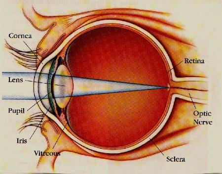
Knowledge of the anatomy of the human eye is important so you may understand the various conditions that can affect your vison and helpful in choosing the appropriate therapy.
Tear film: the true first layer or surface of the eye. Tears are composed of water, salts, oils, mucus, oxygen, and infection fighting compounds. Tears are important for clear vision and the health of the cornea. Lacrimal glands, located under the upper eyelid, make tears.
Cornea: a clear “bubble window” of the eye composed of three layers:
Epithelium: surface cells or clear skin that cover the outer surface
Stroma: transparent connective tissue that provides the shape and form of the cornea
Endothelium: specialized cells that line the back side of the stroma which act as water pumps to keep the stroma dry and clear.
Conjunctiva: a clear coating over the eyeball and backsides of the eyelids
Anterior chamber: a fluid filled space, provides a source of nourishment for the cornea and lens
Iris: a specialized thin colored muscle, controls the amount of light that enters into the eye.
Ciliary body: a circular muscle that supports the lens and controls its shape, allowing the eye to focus at close range
Lens: an “M & M candy” shaped structure, focuses light as it passes towards the retina. Composed of 3 layer =
Capsule: the thin outer clear membrane or shell
Cortex: a clear, thick, protein substance filling the shell
Nucleus: a hard center or core
Zonules: string like structures that connect the ciliary body to the lens and aid in focusing
Vitreous humor: a clear, thick jelly or “egg white” like substance that fills the main eye cavity
Retina: a tissue paper thin layer, contains of millions of light sensitive rod & cone vision cells
Choroid: a vascular spongy layer underneath the retina that provides a blood source to nourish the retina
Sclera: a tough, white shell of the eye made of connective tissue
Optic nerve: a “cable” composed of millions of “wires” from the vision cells, carries vision information to the brain for processing
Optic cup: a small depression or dimple in the optic nerveWe are currently creating content for this section.
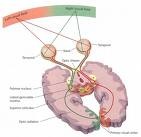
ANATOMY OF THE VISUAL PATHWAY
After capturing light rays, the eyes send this basic information to the visual cortex for detailed processing and eventually the perception of vision is created. The pathway is composed of special sections described below.
Optic nerves -each is composed of millions of nerve fibers that originated in the retina from the rods and cones (vision cells) This "fiber optic cable" transmitts basic visual information from each eye into the brain.
Optic chiasm - a "cross over" point for each optic nerve where vision information from each eye is divided up. The inner retinal visual information (from the outer area of sight) is passed to the opposite side where the outer retinal visual information stays on the same side.
Optic tract - the portion of nerve fiber bundles after the chiasm.
Geniculate body - a relay point where visual information is combined with auditory input for coordination of "looking" towards a sound or noise.
Optic radiations - carry vision information from the geniculate bodies to the occiptial lobes. Fiber orientation becomes compartmentailzed into quadrants. Upper right vision is channed to lower left side of the the occipital lobe
Occipital lobe - Located in the posterior section of the brain. It is the area of final visual processing and eventual perception.


 After capturing light rays, the eyes send this basic information to the visual cortex for detailed processing and eventually the perception of vision is created. The pathway is composed of special sections described below.